Thursday, August 26, 2010

What is Tooth Decay?

Dental cavities are an infection caused by a combination of carbohydrate-containing foods and bacteria that live in our mouths. The bacteria are contained in a film that continuously forms on and around our teeth. We call this film plaque. Although there are many different types of bacteria in our mouths, only a few are associated with cavities. Some of the most common include Streptococcus mutans, Lactobacillus casei and acidophilus, and Actinomyces naeslundii.

  Streptococcus mutans


When these bacteria find carbohydrates, they eat them and produce acid. The exposure to acid causes the pH on the tooth surface to drop. Before eating, the pH in the mouth is about 6.2 to 7.0, slightly more acidic than water. As "sugary foods" and other carbohydrates are eaten, the pH drops. At a pH of 5.2 to 5.5 or below, the acid begins to dissolve the hard enamel that forms the outer coating of our teeth.

As the cavity progresses, it invades the softer dentin directly beneath the enamel, and encroaches on the nerve and blood supply of the tooth contained within the pulp.


Cavities attack the teeth in three ways:
1. Pit & Fissure
2. Smooth surface
3. Root surface

The first is through the pits and fissures, which are grooves that are visible on the top biting surfaces of the back teeth (molars and premolars). The pits and fissures are thin areas of enamel that contain recesses that can trap food and plaque to form a cavity. The cavity starts from a small point of attack, and spreads widely to invade the underlying dentin.


 Decays happen at the the pits and fissures and spread to the biting surface of the teeth

The second route of acid attack is from a smooth surface, which is between, or on the front or back of teeth. In a smooth-surface cavity, the acid must travel through the entire thickness of the enamel. The area of attack is generally wide, and comes to a point or converges as it enters the deeper layers of the tooth.


 Smooth surface decay started at the in-between of teeth
.
The third is the attack started at the root surface of the tooth after it was exposed to the oral cavity. The root is usually exposed due gum recession as a result of periodontitis (gum disease)

Decay on the root surface of the teeth

How Will I Know if I Have a Cavity?
The large majority of cavities are completely painless. This is because the outer enamel has no nerves. It is only when the cavity enters the underlying dentin that the cavity may begin to feel sensitive. The most common cavity symptoms are an increased sensation to cold, sweet foods or beverages. A cavity is often responsible for a broken tooth. The cavity weakens the tooth, especially when it forms under a tooth filling or a tooth cusp, and can easily cause a fracture when biting down.

Patients are sometimes taken off guard when they learn that they have a few cavities but they don't have any symptoms. It is far better to treat a small cavity than to wait until they have symptoms; such as pain. By the time there are symptoms, the cavity may have spread to infect the dental pulp, necessitating a root canal procedure or a tooth extraction to eliminate the infection. Always remember that most dental problems are insidious -- that is, they sneak up on you. Regular dental exams, at least twice a year, will greatly reduce the likelihood that a dental cavity will go undetected and spread, causing toothache pain and infecting the dental pulp.

The decay has spread into the dental pulp causing pain

How Do Dentists Detect Cavities?
Cavities are detected a number of ways. The most common are clinical (hands-on) and radiographic (X-ray) examinations. During a clinical exam, the dentist uses a handheld instrument called an explorer to probe the tooth surface for cavities. If the explorer "catches," it means the instrument has found a weak, acid damaged part of the tooth -- a dental cavity. Dentists can also use a visual examination to detect cavities. Teeth that are discolored (usually brown or black), can sometimes indicate a dental cavity.

 Regular dental examination is important to prevent tooth decay

 Bite-wing radiograph is good to detect interproximal (in-between) caries

Dental X-rays, especially check-up or bitewing X-rays, are very useful in finding cavities that are wedged between teeth, or under the gum line. These "hidden" cavities are difficult or impossible to detect visually or with the explorer. In some cases, none of these methods are adequate, and a dentist must use a special disclosing solution to diagnose a suspicious area on a tooth.

Are Some People at More Risk for Developing Cavities?
People who have reduced saliva flow due to diseases such a Sjogren Syndrome; dysfunction of their salivary glands; have undergone cancer chemotherapy or radiation; and who smoke are more likely to develop cavities. Saliva is important in fighting cavities because it can rinse away plaque and food debris, and help neutralize acid. People who have limited manual dexterity and have difficulty removing plaque from their teeth may also have a higher risk of forming cavities. Some people have naturally lower oral pH, which makes them more likely to have cavities.

How Can I Prevent Cavities?
The easiest way to prevent cavities is by brushing your teeth and removing plaque at least three times a day, especially after eating and before bed. Flossing at least once a day is important to remove plaque between your teeth. You should brush with a soft-bristled toothbrush, and angle the bristles about 45 degrees toward the gum line. Brush for about the length of one song on the radio (three minutes). It's a good idea to ask your dentist or hygienist to help you with proper brushing methods.


 Blushing and Flossing teeth are to do it daily to stop caries

Reducing the amount and frequency of eating sugary foods can reduce the risk of forming cavities. If you are going to drink a can of sweetened soda, for instance, it is better to drink it in one sitting, than sip it throughout the day. Better yet, drink it through a straw in one sitting, to bypass the teeth altogether. Getting to the dentist at least twice a year is critical for examinations and professional dental cleanings.

 Reduce high sugar food can reduce dental cavity significantly

To reduce the incidence of cavities, use toothpaste and mouthwash that contains fluoride. Fluoride is a compound that is added to most tap water supplies, toothpastes, and mouth rinses to reduce cavities. Fluoride becomes incorporated into our teeth as they develop and makes them more resistant to decay. After our teeth are formed, fluoride can reverse the progress of early cavities, and sometimes prevent the need for corrective dental treatments.
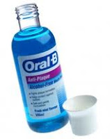
Mouthwash with fluoride

The recent drop in the number of cavities is largely due to the addition of fluoride to our drinking water. Mass water fluoridation is the most cost-effective measure available to reduce the incidence of tooth decay. The Environmental Protection Agency has determined that the acceptable tap water concentration for fluoride is 0.7 to 1.2 parts per million.


A dental procedure called sealants can also help reduce cavities on the top and sides of back teeth (occlusal, buccal and lingual surfaces). A sealant is a white resin material that blankets the tooth, protecting the vulnerable pits and fissures of the tooth. Sealants are routinely placed on children's teeth to prevent cavities on their newly developing molars. The use of sealants to prevent cavities is also a cost-effective way to reduce the incidence of cavities on adults as well. Sealants are generally not used on teeth that already have fillings.

 Fissure Sealant

People who have a dry mouth are at risk for developing cavities, and can have their dentist prescribe artificial saliva and mouth moisturizers, as well as recommend chewing sugarless gum to stimulate saliva production. Finally, an antiseptic mouthwash containing chlorhexidine gluconate such as Chlohexxa or Oradex can also be useful in killing bacteria associated with dental caries.

What should I do if I have tooth decay?
You should go the to dental clinic as soon as possible. Early or small decay is easily to treat. Usually a small filling will do. However if it is large cavity, then a larger filling is required provided there is no pain. In cases where the tooth is painful (eg. pain on biting, disturb sleep), then root canal treatment or extraction is required to stop the infection.
 
Small filling
Filling can be silver (amalgam) or white (composite).


Large Filling
Usually required
Comparison within big and small filling:

      Small Filling                           vs.       Large filling
  • Less pain during filling                    More pain (because lager & deeper cavity)  
  • More aesthetic                               Less aesthetic
  • More lasting and durable                Less durable
  • Cheaper                                        More expensive (more filling material)
Crown
Or tooth capping of is a procedure to created back function, aesthetic as well as protection to a severely damaged tooth. It is usually made of porcelain fused with metal or a full porcelain material. Crown is durable and more lasting compared to a large filling.


Root canal treatment (RCT)
RCT is required when infection from caries has spread to the pulp of a tooth. The tooth is usually painful on chewing and sometimes disturb sleep. The purpose of this treatment  is to preserve the tooth by removing the dead and infected pulp leaving the tooth bacteria free.

After RCT, the tooth can be restored with filling or a corwn. If there is a lot of tooth structure loss, the tooth should be protected with a crown. 


Extraction
Tooth extraction in another way to stop infection. However, this method is commenced  if patient don't want to keep the tooth anymore. Patient have to understand the consequent of removing the tooth

      Root canal treatment              vs.        Tooth extraction

  • Tooth preserved                             Tooth removed
  • Difficult (esp molar tooth)                Simple & fast
  • Expensive                                       Cheaper than RCT
  • Few visits                                       One visit
  • Lesser problems in future                 More problems in futures

For more info on  problems with missing tooth/teeth and how to overcome them, click here


...

Monday, August 23, 2010

Problems with missing teeth

As soon as a tooth is being removed, we have to think of the options available for replacing that missing tooth. Most of us don't really know the consequences or future problems that are going to be faced by us resulting from tooth extraction.

These are some of them:
Chewing efficiency decrease
The most common problem patient face immediately after extraction it lost of chewing efficiency. He will tends to chew more on the opposite side. If the extraction involved multiple teeth, then patient will have to switch to soft diet which might result in poor digestion and malnourished. Therefore replacement of missing teeth is crucial to regain back patient's normal chewing.

Over eruption of opposite tooth
Commonly happen after extraction of the lower molar causing the upper molar to erupt further downwards. This will result in food stagnant in the interproximal (in-between of the teeth) area making the teeth easily develop dental caries.

Migration of neighbour tooth
The neighbour teeth will migrate to the extraction site as soon as the tooth was remove. Usually, the patient will begin to notice it after a year or more. In some delayed cases, tooth extraction causes the front teeth to have multiple gaping which resulted in poor aesthetic. Both migration and supra-eruption of teeth will make restoration or replacement of the missing tooth difficult.

Bone lost
Alveolar bone lost significantly after missing teeth. As the result, the upper lip looks flatten due to lost of support from the anterior alveolus bone. Lost of bone also makes implant insertion difficult which might required bone harvesting before implant insertion.

Attrition of the remaining teeth
When patient loose most of his back teeth, he will try to use his front teeth to grind food and eventually all the front teeth will look much shorter due to attrition from chewing. Attrition also will make the teeth become sensitive to cold and sweet (dentinal hypersensitivity).

Over-closure of the mouth

As the result from attrition of front teeth and lost of posterior chewing, patient tends to over close his jaw. This will make his face shorter and his lip looks thin and easily get fungal infection at the corner of the mouth

Traumatic occlusion and Jaw joint dysfunction
Missing teeth will cause parafunction (abnormal) chewing. For example, when the back teeth are missing, the front teeth will be used for grind. This abnormal force will be exerted to the remaining teeth causing bone resorption around the teeth and lead to gum problem and loosening of teeth. Heavy and abnormal chew will also causing the TMJ (jaw) joint pain.
Due to abnormal function, the remaining teeth have a very high chance of fracture.

Conclusion
Delay in restoring missing teeth will result in:
1. Loss of chewing
2. Difficult in restoration/replacing missing teeth
3. Poor dental aesthetic - short teeth, teeth gaping, deep bite
4. Poor facial aesthetic - over closure, short face, chin protrusion
4. Prone to dental decay, gum problem, tooth fracture, dentine hypersensitivity
5. And finally, making you look older....


Treatment Options for Replacing Missing Tooth/Teeth
Option 1: Denture


Advantages
1. Cheap
2. Complexity: Simple
3. No need needle injection
4. No need surgery (No pain)
5. Maintenance: Easy to take care
6. Treatment duration: Short 1 - 3 weeks
7. Easy to adjust, repair

Disadvantages
1. Feel like not real (fake teeth) - can be removed from the mouth
2. Uncomfortable - Big and bulky
3. Palate coverage - less taste when eating
4. Lower ridge coverage - no space for the tongue
5. Teeth is made of plastic - easily worn off & stained
6. Easily trap food
7. For one missing tooth - wearer is not willing to wear it, very uncomfortable
8. Chewing food not the same as the natural teeth
9. Easily break

Option 2: Bridge


Advantages
1. Feel like real teeth - cannot be removed
2. Highly aesthetic - Look like natural teeth (with full porcelain), multiple shade to select
3. Very comfortable - No palate or lower ridge coverage
4. Teeth is made of porcelain - strong, durable
5. Good for missing one or a few teeth
6. Can correct the abutment teeth to a desirable shape and position
7. Chewing food almost as real as natural teeth
8. No need surgery

Disadvantages
1. Price: Moderate
2. Complexity: Simple to complex
3. Required needle injection
4. Involved neighbour teeth - the neighbour teeth required to be ground for support
5. Easily trap food under the bridge
6. Maintenance: Difficult (Required to floss under the bridge)
7. Treatment duration: Medium 2 - 3 weeks
8. No suitable for cases such as free end saddle, fully or near to edentulous

Option 3: Dental Implant

Advantages
1. Feel like real teeth - porcelain is sitting on the implant
2. Highly aesthetic - Look like natural teeth (with full porcelain), multiple shade to select
3. Very comfortable - No palate or lower ridge coverage
4. Teeth is made of porcelain - strong, durable
5. Does not involve the neighbour teeth
6. Trap food: very minimal (just like natural teeth)
7. Maintenance: simple to take care (just like natural teeth)
8. Can replace missing teeth in edentulous and free end saddle
9. Chewing food almost as real as natural teeth

Disadvantages
1. Price: Expensive
2. Complexity: Complex - required good planning
3. Duration of treatment: Long 3 - 6 months
4. Required needle injection
5. Required to undergo surgery (maybe required second or third surgery)
6. Required sinus augmentation, bone harvesting if not enough bone height for implant insertion (additional cost, additional surgery....additional pain)
7. Higher risk of failure in smoker, diabetic and medically compromised patient
8. Risk of injuring other structure during implant insertion: ID nerve, antrum

Click here to know more about implant


...

Tuesday, August 17, 2010

Dental Implant

What is dental implant?
Dental implant is an artificial root of the tooth used in dentistry to support restorations that resemble a tooth or group of teeth.
There are a lot of implant system in current market with different shape and size


All dental implants today (21st century) are root-form and they are placed inside the bone (endosseous implant). Prior to the advent of root-form endosseous implants, most implants were either blade endosseous implants, in that the shape of the metal piece placed within the bone resembled a flat blade, or subperiosteal implants, in which a framework was constructed

Implant is used to support a crown (for the case of missing a single tooth) or they can be used to support bridge or denture (for the case of multiple missing teeth)


  Composition of an Implant
A typical implant consists of a titanium screw (resembling a tooth root) with a roughened or smooth surface. The majority of dental implants are made out of commercially pure titanium, which is available in 4 grades depending upon the amount of carbon and iron contained. More recently grade 5 titanium has increased in use which offers better tensile strength and fracture resistance.  Implant surfaces may be modified by plasma spraying, anodizing, etching or sandblasting to increase the surface area and the integration potential of the implant.


Anatomy of Implant
Implant/Fixture - titanium material for osteo-integration with the surrounding bone.
Abutment - structure which connects the implant to the crown and it is situated in the gum area.
Crown - Usually porcelain fused to metal material that can be seen in the mouth.

Who should perform the Implant placement?
Implant surgery may be performed as an outpatient under general anesthesia, oral conscious sedation, nitrous oxide sedation, intravenous sedation or under local anesthesia by trained and certified clinicians including general dentists, oral & maxillofacial surgeons, prosthodontists, and periodontists.


Surgical procedure
Surgical planning
Prior to commencement of surgery, careful and detailed planning is required. Two-dimensional radiographs, such as orthopantomographs (OPG) or periapicals radiograph are often taken prior to the surgery. They are used to identify vital structures (such as the inferior alveolar nerve or the sinus), as well as the shape and dimensions of the bone to properly orient the implants for the most predictable outcome. In some instances, a Cone beam tomogram (CBT) or CT scan will also be obtained.


Jaw X-ray -- Dental Panaromic Tomogram (or OPG)
 
Cone Beam Images of the jaw bone
Basic procedure
 In its most basic form the placement of an osseointegrated implant requires a preparation into the bone using either hand osteotomes or precision drills with highly regulated speed to prevent burning or pressure necrosis of the bone. After a variable amount of time to allow the bone to grow on to the surface of the implant (osseointegration) a tooth or teeth can be placed on the implant. The amount of time required to place an implant will vary depending on the experience of the surgeon, quality and quantity of the bone and the difficulty of the individual situation (usually between 30 minutes and 2 hours).

Preparation of recipient bone for implant insertion

Surgical procedure
An incision is made over the crest (highest point of the gum ridge) of the site where the implant is to be placed. The gum (which is referred to as a 'flap') was raised  to exposed the recipient bone. Then, a pilot hole is bored into the bone, taking care to avoid the vital structures. Drilling into jawbone usually occurs in several separate steps. The pilot hole is expanded by using progressively wider drills (typically between three and seven successive drilling steps, depending on implant width and length). Care is taken not to damage the bone cells by overheating. A cooling saline or water spray keeps the temperature of the bone to below 47 degrees Celsius.

Implant insertion into recipient bone

The implant screw can be self-tapping, and is screwed into place at a precise torque so as not to overload the surrounding bone (overloaded bone can die, a condition called osteonecrosis, which may lead to failure of the implant to fully integrate or bond with the jawbone). Typically in most implant systems, the osteotomy or drilled hole is about 1mm deeper than the implant being placed, due to the shape of the drill tip. Surgeons must take the added length into consideration when drilling in the vicinity of vital structures.


Before and after implant insertion on the upper right area


Healing time
Practitioners usually allow 2–6 months for healing. If the implant is loaded too soon, it is possible that the implant may move which results in failure. The subsequent time to heal, possibly graft and eventually place a new implant may take up to eighteen months. For this reason many are reluctant to push the envelope for healing.
Wound healing. Left pic - immediate after surgery, Right pic - 7 days after surgery in one-stage surgery.


One-stage or two-stage surgery?
When an implant is placed with a 'healing abutment', which comes through the mucosa, it is called the One-Stage surgery. (Picture above is one-stage surgery where the healing abutment - green and purple screw can be seen on the gum surface)

When an implant is placed with  a 'cover screw' which is flush with the surface of the dental implant and it is covered with the mucosa/gum, this surgery is the Two-stage surgery. A second surgery is needed 3 months later to exposed the cover screw and to be placed the healing abutment.

Two-stage surgery is sometimes chosen when a concurrent bone graft is placed or surgery on the mucosa may be required for esthetic reasons. Some implants are one piece so that no healing abutment is required.
In carefully selected cases patients can be implanted and restored in a single surgery, in a procedure labeled "Immediate Placement". In such cases a provisional prosthetic tooth or crown is shaped to avoid the force of the bite transferring to the implant while it integrates with the bone.

Surgical timing
There are different approaches to place dental implants after tooth extraction. The approaches are:
Immediate post-extraction implant placement.
Delayed immediate post-extraction implant placement (2 weeks to 3 months after extraction).
Late implantation (3 months or more after tooth extraction).


Restoration Procedure
For missing single tooth
Implants can be made to replace missing tooth or teeth. If an implant is used to replace one missing tooth, it is  implant-supported crown.

Implant-supported crown is used to replaced a missing tooth

Multiple missing tooth
Implant-supported crown can be used to replaced multiple missing tooth. Every missing teeth will be replace with implants


If in cases where the bone is too narrow and not suitable for implant placement then implant-supported bridge will be used to avoid those unsuitable area.

Implant-supported bridge also can be used to reduce implant cost and surgical time!!

Impression taking
After the implant in well osteo-integrated with the surrounding bone, construction of the outer part -- the crown/tooth procedure can be started. Usually it started with impression taking after the gum heals around the healing abutment.

 Gum healing after a week on healing abutment in two-stage surgery


Impression is used to make a duplication of the gum and the implant position where the laboratory technician can fabricate crown/bridge outside patient's mouth.

The impression transfer posts are inserted on the implant fixtures 

And impression of the gum and implant is taken with silicone material


Crown/Bridge fabrication
In the dental laboratory, porcelain fused to metal crowns or bridges are fabricated. Usually it takes about 2-3weeks to be done.

A model with implant is made from the silicone impression.

On the model, crowns are fabricated and ready to be fit in patient mouth!!

Another case with two implant-supported bridges


Restoration crown/bridge in the mouth
There are basically 2 types of ways by which the crowns/bridges that can be attached to the implant:
Screwed retained - The crowns/bridges are retained in the mouth with screw that screw into the abutment. The advantage of using this way is easy to be removed when ever need (eg. if the crown break or the abutment become loosen). However, the aesthetic will be compromised. Usually screwed retained method is used in posterior region where aesthetic is not an issue or in straight abutment type.
Cemented retained - The crowns/bridges are cemented onto the abutment with cement such as temp-bond, GIC etc. The advantages is very good aesthetic. Usually used in the anterior region or in angle abutment type.


Below are the procedures of installing the final restoration of a screw-retained type:

 After removing the healing abutment, the implant 'hole' will be assessed

The abutments are screwed into the implants with force 15Ncm

Then, the crowns are fit onto the abutments and the colour, shape and occlusion will be assessed

After that,  crowns will be screw onto the abutments with force of 15Ncm

 The hole at the palatal surface will be covered with filling material

Final result!!


...